Ocular Disease
Ocular Disease
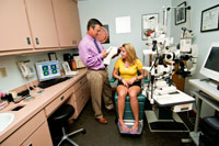
The Wilcox Eye Center (WEC) does not believe there is ever a “routine eye exam.” We respect that sight is a precious gift and balance “routine vision needs” (wellness exams, glasses and contacts lenses) with a consistently suspicious look out for the unsuspected, overt or occult, ocular disease. Dr. Peter E. Wilcox is very experienced in the diagnosis, treatment and management of ocular disease. Dr. Wilcox chooses to lecture to national and international audiences and spends the time and study to be published in the Optometric literature. Please read his Curriculum Vitae and examine a sampling of his educational efforts, lectures and publications. In addition, Dr Wilcox is a consultant to contact lens manufacturers.
In the rapidly-changing healthcare field it is not uncommon for a Dr-patient experience to be brief, sterile, uncomfortably compartmentalized and impersonal. It is the intention of the WEC to balance timely delivery of care while generously investing in appropriate testing, thought and communication of diagnoses/options to every patient. These philosophies are practiced in each step of the patient experience at the Wilcox Eye Center; from the phone conversations, face-to-face clinical and optical experiences and the time spent behind the scenes to continue/complete your care.
The correct answer learned in first year Optometry/Medical school to the question: “When is the Case History over?” is; “The Case history NEVER ends!” So, what does this mean to our patients? It should give patients/parents comfort that amidst the typically jovial and family-oriented delivery of care, we are always thinking and testing globally for the patient’s best interest.
Another great virtue of the WEC is the detailed communications to the other health care providers. It is not uncommon for Dr Wilcox to immediately, electronically communicate with another specialist or PCP or allied health provider while the patient sits in the chair or shortly thereafter. The communication or referral typically includes test results, attachments, annotations and other clarifying information to assure the message is understood. Please read the friendly e-Publication: “It’s Kind of Like… The Art of Patient Communication” published in 2/13. It reveals the acumen Dr Wilcox utilizes to assure that the recipient of communications truly understands the What’s and Why’s of a given circumstance.
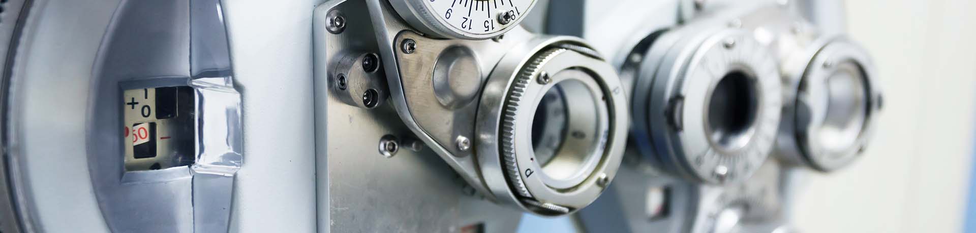
Though we optimize the age-old instruments most familiar with during a typical ‘eye exam’ (pen light, phoropter (the “Which is better 1 or 2?” instrument with lots of lenses), slit lamp, keratometer, color vision charts, depth perception testing. Let’s review some of the non-routine instruments/technologies we utilize. Note, all of our technologies are networked to assist in patient flow and information retrieval…
Visual Field Analysis:
We use a screening visual field instrument called the FDT and a more detailed automated visual field unit.
FDT: This instrument is used in pretesting to quickly analyze the patient’s visual field. It is designed to be efficient and timely. The results are typically uneventful however at times there are losses registered during the test. Any losses are evaluated and cross referenced to other confirmatory same-day visual field testing. A decision is then made to engage the more formal field unit.
Negative diagnoses made with this unit range from the relative insignificance of poor test-taking to the life-saving diagnosis of undiagnosed brain tumor.
Humphrey’s Visual Field Analyzer: This instrument yields a much more detailed understanding of the patient’s visual field wellness. It is routinely used in the management of our glaucoma patients as well as those with various macular and optic nerve/radiations diseases, potential drug toxicities and with those who complaint of headaches – which may be secondary to intracranial complications.
Topographer:
A corneal topographer is used to map the surface curvatures of the clear dome of the eye, the cornea. The information allows us to understand the wellness of the cornea. There are many versions of healthy corneas. They come in 3 basic curvatures steep, average and flat. You can toss in astigmatism which is a difference in the curvatures in the cornea when looking at different diameter cords. Knowing the specifics of the surface of the cornea is of upmost importance in knowing how and why a patient sees and experiences vision the way they do. For example, two different patients with the SAME prescription of -1.00 diopters of sphere and 1.50 diopters of cylinder at a certain axis may have very different corneal topographies and subsequently totally different net vision perceptions. Not knowing a patients’ topography can cause a great deal of guessing and typically wrong assumptions by the eye doctor. We routinely perform topography so not to be in such a deficit.
The WEC uses two topographers given their continual daily use. The more significant fact is they are the Scout Topographer from EyeQuip®. The Scout topographer is relatively unique because a design software program called Wave® is linked which allows the creation of pre-corneal lenses (fancy contacts) to achieve numerous patient needs and goals. The Wave lenses designed by Dr. Wilcox are/for:
Single Vision and Multifocal (to wear all day)
- Corneal = diameters less than the corneal diameter
- Corneal-Scleral = diameters slightly larger than the cornea which creates great comfort and assists with certain corneal or refractive needs.
- Semi-Scleral = Larger diameter and are used for healthy eyes but more importantly unhealthy eyes (Post-refractive surgery, corneal transplants, severe dry eye, keratoconus and many/any other corneal diseases)
- Scleral Lenses = for the unhealthy eyes. Some of the disease cases listed above and those eyes in a more significant case of compromise.
- Myopic Corneal Molding: the creation of a topography-based lens for the nighttime wear which reshapes the cornea so the patient can see great all day WITH NOTHING IN THEIR EYES. It also stops-to-slows the progression of nearsightedness in kids.
- Hyperopic Corneal Molding: the creation of a topography-based lens for the farsighted eye or the over-40-something who needs reading glasses
The topography-based lenses are superior to most other choices as they are based on thousands and thousands of data points unique to the ‘finger print’ of the individual cornea and not a handful of called-in design characteristics. www.wavecontaclenses.com
Understanding Corneal Topography Read one of Dr. Wilcox’s publications pertaining to the value of topography and topography based lenses.
Ocular Coherence Tomography (OCT):
There is so much to say about the extraordinary value that the OCT brings to the World of Eye Care. This technology was brought to eye doctors in the study of the retina and optic nerve. As always, technology improves and grows. The WEC invested in the second generation RTVue SD-OCT. OptoVue is the manufacturer which leads the industry while allowing the evaluation of the front of the eye. Almost a full step ahead of the manufacturer’s intent, Dr. Wilcox knew that this leap in visualization would allow him to evaluate the performance of contact lenses on the eye. He was the first in the State of Virginia to bring the Opto-Vue SD-OCT into practice.
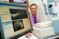
This technology allows visualization, analysis monitoring and management of ocular disease in ways simply unimagined a decade ago. As there is too much to list, papers and links will serve best to educate. Dr Wilcox uses the RTVue SD-OCT to evaluate the fit of various contact lenses. His study and thoughtful application of innovative design and fitting logic has assisted in making him relevant in the arena of specialty gas permeable contact lenses. www.optovue.com
Retinal Photography
Retinal photography serves two purposes: (1) To document a normal retina/optic nerve = fundus. This is immediately diagnostic and serves as a perfect benchmark for any possible future changes. (2) To capture newly yet-undiagnosed diseases or conditions and/or to electronically monitor known eye diseases. The WEC invested in the Cannon Synemed non-mydriatic camera. www.synemed.com/nonmydriatic.html
Slit Lamp Photography:
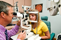
Slit lamp photography is NOT limited to external photography. Anything you can see in the slit lamp can be captured with digital imaging; from the retinal to the cornea to lenses fit on the eye and external ocular/skin conditions.
The same relative purposes for retinal photography are applied to slit lamp photography. The WEC has three Tel-Screen networked slit lamp camera systems. Images are captured for patient education, chart documentation and inter-professional referrals and communications. www.telscreen.com
A Pledge from Dr. Wilcox
It is the mission of the Wilcox Eye Center to exceed each patient’s expectations by providing the most comprehensive and highest quality vision care services and products available.
We will deliver this optimal level of eye care by remaining on the cutting edge through active research and continued education while investing in proven state-of-the-art technology.
We will treat each patient with respect and create an enduring relationship based on trust and honest communication.
Our Services At-a-Glance
Address:
Square Suite 1
2652 George Washington Memorial Highway
Hayes, VA 23072
Email:
Phone:
804-642-9800